
Advent, Advent, ein Lichtlein brennt. Erst eins, dann zwei, dann drei …
(Advent, Advent, a little light is shining. First one, then two, then three …)
Christmas is approaching fast and we are already lighting our third candle on our scientific Advent wreath at CALA! Each week of the Advent season, our DOLPHIN group led by Andreas Döpp is lighting a special candle to showcase techniques normally applied to their experiments at CALA. In the context of laser-plasma interactions, the group develops diagnostics to study plasma – the “fourth state of matter” – in high-power laser experiments. The Advent project was carried out by Matilde Nunes, Johannes Altmann and Marguerite Dion.
Third candle: Analyzing emission lines with a hyperspectral camera
Last week, the researchers used a hyperspectral camera to create a temperature map of a candle flame. This week, they used the same camera – and in fact, the same measurements – to identify specific elements within the flame and where they are located.
As we learned last week, a hyperspectral camera captures a full spectrum of light at every point of the image, unlike a traditional camera that only records three color channels (red, green and blue). Since each chemical element emits light at its own characteristic wavelengths – essentially like a unique fingerprint – this full spectrum allows the researchers to pinpoint the presence of specific elements.
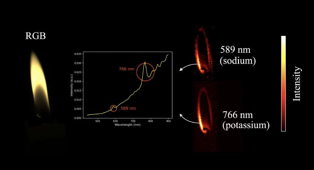
The flame contains clear signatures of sodium and potassium, which likely originate from trace amounts of these elements in the candle wax. By analyzing the full spectrum at each pixel, the researchers could map the emission intensities for both elements across the entire candle flame.
Both maps show that the emission is stronger in the outer regions of the flame. These zones are hotter and contain more complete combustion. Therefore, more atoms are excited, and the intensity of the emission lines is higher. The inner part of the flame, where unburned vapor is present, is cooler and therefore emits less strongly.
Hyperspectral imaging is a powerful tool that usually comes into play when three color channels alone are not enough to distinguish between different light spectra. This is particularly useful for fields like agriculture and biomedical imaging, where the technique can for example be used to diagnose plant diseases or to detect skin diseases.
Hyperspectral imaging techniques are also used in laser diagnostics, which is closely connected to high power laser physics. At the DOLPHIN group, researchers combine optics with data driven methods to develop new tools for hyperspectral imaging. A commercial hyperspectral camera, like the one that captured this week’s candle image, can be used to validate new techniques or to capture datasets for the data driven methods.
Next week, we will reveal the final candle and method on our scientific Advent wreath!
Pictures: Advent candles: AI-generated / Nina Beier; Measurements: Matilde Nunes, Johannes Altmann and Marguerite Dion


